
Ureters
Each ureter is a small tube, about 25 cm long, that carries urine from the renal pelvis to the urinary bladder. It descends from the renal pelvis, along the posterior abdominal wall, which is behind the parietal peritoneum, and enters the urinary bladder on the posterior inferior surface.
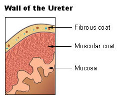
The wall of the ureter consists of three layers. The outer layer, the fibrous coat, is a supporting layer of fibrous connective tissue. The middle layer, the muscular coat, consists of the inner circular and outer longitudinal smooth muscle. The main function of this layer is peristalsis: to propel the urine. The inner layer, the mucosa, is transitional epithelium that is continuous with the lining of the renal pelvis and the urinary bladder. This layer secretes mucus, which coats and protects the surface of the cells.